|
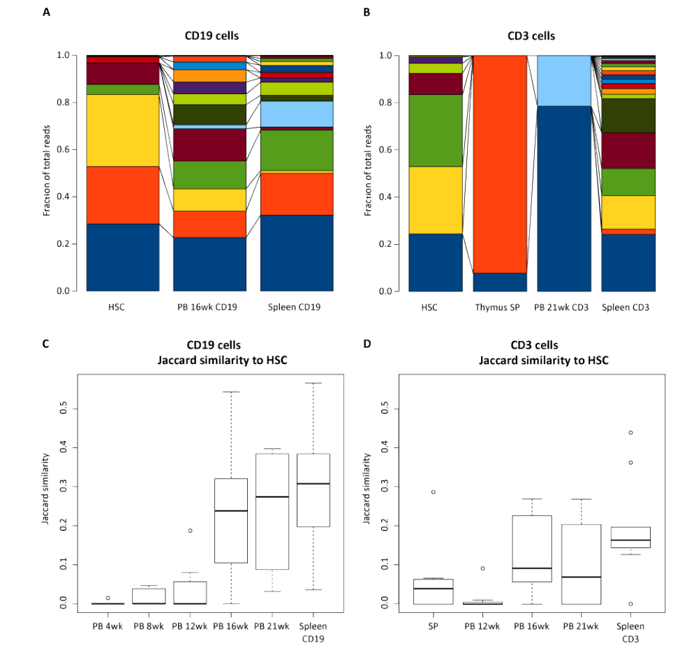
| Figure 2: Different clonal contribution in T vs B cell clones. Data from 9 different NSG mice transplanted with barcoded purified human HSCs. The individual clones are represented by different colors, with the full bar representing 100% of that population. (A) For B cells, HSC, peripheral blood at 16 wk post-transplant and spleen 16 weeks post-transplant are shown for a representative animal. (B) For T cells, HSC, single positive thymocytes, peripheral blood and spleen are shown at 21 weeks. The Jaccard index was calculated between HSC and the indicated samples obtained in 9 mice in the CD19 (C) or CD3 (D) sorted samples. The diversity in B cells is greater and arising from different clones compared to T cells |