 |
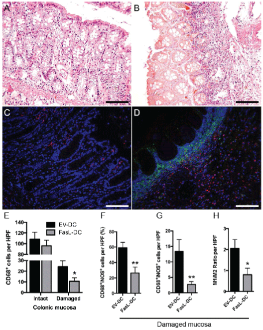
| Figure 5: Adoptive transfer of dendritic cells expressing FasL (FasLDCs) decreases the proportion of colonic proinflammatory macrophages. Representative micrographs of hematoxylin and eosin staining depicting the histology of intact (A) and damaged (B) colonic mucosa from colitic rats (400x). Representative micrographs of double immunofluorescent staining for CD68 (red) and inducible nitric oxide synthase (iNOS, green) of intact (C) and damaged (D) colonic mucosa from colitic rats (400x). (E) Number of CD68+ cells per high-power field (HPF, 400x) of colonic tissue from rats treated with FasL-DCs or dendritic cells transfected with an empty vector (EV-DCs). (F) quantification of CD68+iNOS+ cells per HPF of colonic tissue from rats treated with FasL-DCs or EVDCs, expressed as a percentage of total CD68+ cells. (G) Number of CD68+iNOS+ cells per HPF of colonic tissue from rats treated with FasL-DCs or EV-DCs. (H) M1/M2 ratio (calculated CD68+iNOS+/CD68+iNOS-) per HPF of colonic tissue from rats treated with FasL-DCs or EV-DCs. n=14-15 HPFs for intact mucosa, 15-17 HPFs for damaged mucosa. * p<0.05, ** p<0.01 vs EV-DC group. Scale bars = 100 μm. |