|
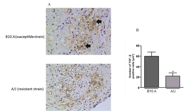
| Figure 5: (A) Expression of TGF-β, detected by immunohistochemichal analysis, in the granulomatous lesions of omentum of susceptible (B10.A) and resistant (A/J) mice, 120 days after ip infection with the virulent Pb18 P. brasiliensis isolate. Arrows point to the TGF-β positive yeast cells in the susceptible mice. The figures are representative of at least three experiments performed on different experimental days. Magnification: 400x. (B) The graphic corresponds to the quantitative analysis of selected microscopic fields of lesions from susceptible (B10.A) and resistant (A/J) mice in terms of TGF-β positivity. Results are expressed in total pixels and represent the mean ± SEM for at least five animals. ** p<0.01. |