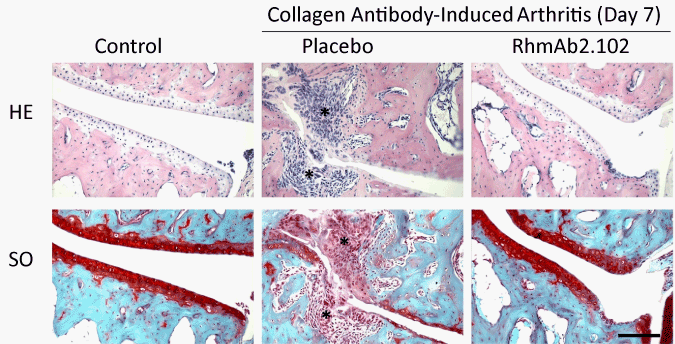
On day 0, 3.2 mg anti-collagen-II antibody mix was injected i.p. in DBA/J1 mice to induce CAIA, followed by an i.p. injection containing 25 μg LPS on day 3, and RhmAb2.102 or placebo i.p. injection on day 7 (1 mg/mouse). Paraffin sections were made from right hind paws and consecutive sections were stained with either Haematoxylin-Eosin staining (HE) or Safranin O staining (SO). Control mice did not receive CAIA antibodies. Blue and red staining indicates bone and cartilage respectively. * indicates inflammatory infiltrate. Bar represents 100 μm.