|
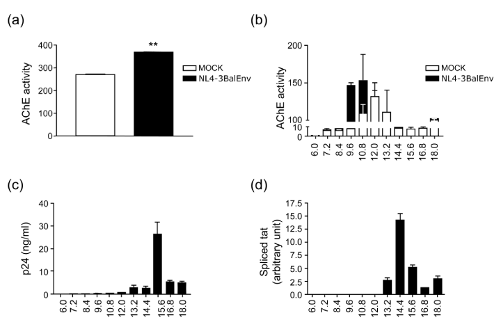
| Figure 3: DCs infected with HIV-1 release exosomes and virions having distinctive markers and sedimentation velocities. DCs were incubated for 1h with mock viral suspension (open bars) or with exosome depleted suspension of NL4-3Balenv produced on PBMCs (black bars) and then cultured for an additional 48h. Cellfree supernatants were subjected to differential centrifugations to pellet exosomes and HIV-1. Exosomes were then separated from virions using Optiprep gradient separation. Exosomes in the 100,000xg pellet (a) and in each individual Optiprep fraction (b) were quantified by measuring AChE activity. The virions were quantified using an anti-p24gag ELISA (c). Data correspond to the mean ± SEM of triplicate samples from one donor and these results are representative of three independent donors. ** P < 0.01. The infectivity of viruses present in each gradient fraction was evaluated by means of real-time PCR targeting the spliced TAT gene in autologous T CD4+ lymphocytes (d). |