|
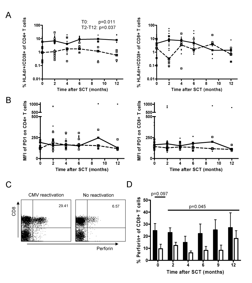
| Figure 2: T-cell activaton, PD-1 and perforin-expression in patients with and without CMV-reactivation. Percentage of HLAdr+/CD38+ (A) and MFI of PD-1 (B) in CD4+ and CD8+ T cells of patients with (black square; median straight line) and without (open square; median dashed line) CMV-reactivation. C. Representative example of perforin-expression by CD8+ T cells in a patient with and without CMV-reactivation, two months after SCT. Percentages of perforin+ T cells within the CD8+ T cells are indicated in the upper right corner. D. Mean (and S.E.M.) percentage of perforin-expression in patients with (black bars) and without (white bars) CMV-reactivation. |