|
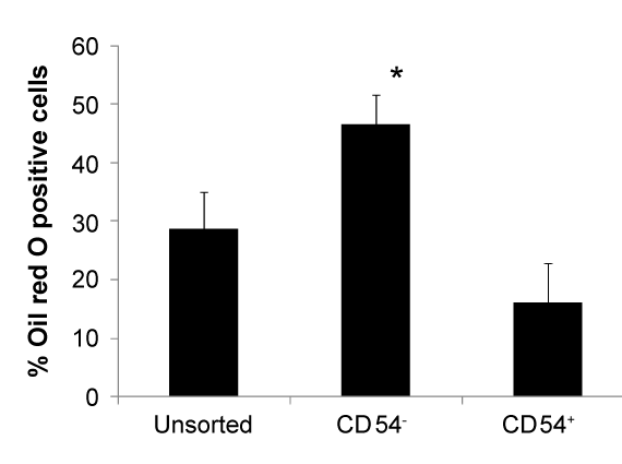
| Figure 6: Quantitative evaluation of adipogenic differentiation of CD54-, CD54+, and unsorted AMSCs. Histological staining of lipid droplets was analyzed under light microscopy. The percentage of oil red O stained cells was calculated as the number of oil red O positive cells divided by the total number of cells as indicated by hematoxylin nuclear staining in 3 different fields (*p<0.05). |