|
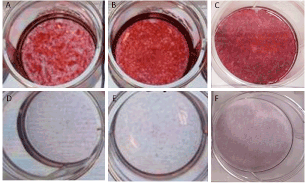
| Figure 7: Osteogenic differentiation of CD54-, CD54+, and unsorted AMSCs. CD 54+ (A and D), CD54- (B and E), and unsorted (C and F) AMSCs were cultured in osteogenic induction medium (A, B, and C) or normal DMEM medium (D, E, and F). After 21 days, alizarin red staining was used to illustrate calcification of mineralized extracellular matrix formed in the induced cells. |