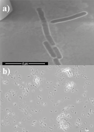
Figure 1:
Bacillus horneckiae
strain APA micrographs after 24h of incubation at optimal growth conditions. 1a) electron microscopy of vegetative cells and 1b) vegetative and sporulating cells under phase contrast microscopy.