|
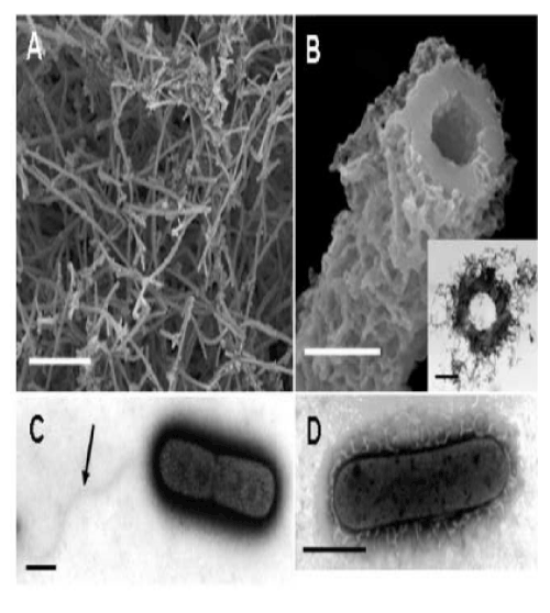
| Figure 1: Aged sheaths in 2-week culture and bacterial cells in 1-day culture. (A) SEM image of numerous tubular sheaths formed on a Fe plate surface in 2-week culture. (B) Enlarged SEM image of an aged sheath and TEM image of its cross-section (inset). (C) Negative staining image of a dividing cell with a monotrichous polar flagellum (arrow) in 3-day culture. (D) Negative staining image of a cell secreting materials of electron-lucent fibrous appearance in 3-day culture. Scale bar: (A) 50 µm, (B-D) 1 µm. |