|
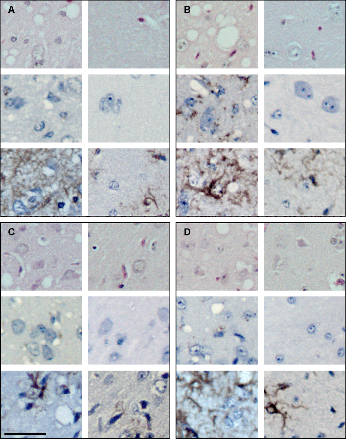
| Figure 4: Histological changes in the hippocampus (A), thalamus (B), cortex (C), and hypothalamus (D) of scrapie-infected (first and third columns) and uninfected (second and fourth columns) hamsters, 61 days post injection. Haematoxylin and eosin staining for vacuoles: first and fourth rows; immunohistochemistry for PrP (second and fifth rows), and GFAP (third and sixth rows). Size bar = 50 microns. |