 |
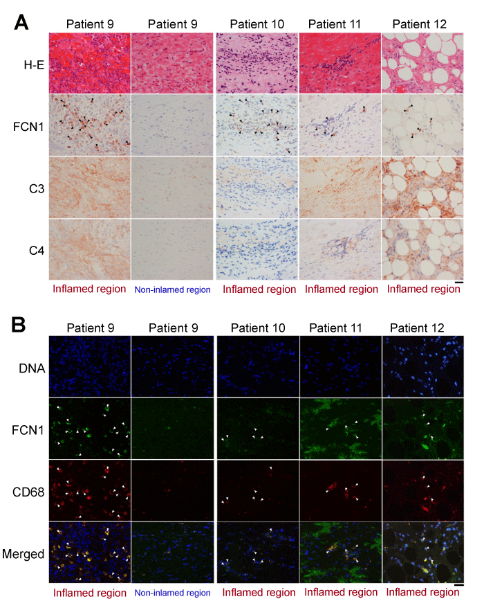
| Figure 6: Immunodetection of FCN1, C3 and C4 in the aortae of patients with TA. (A) Four serial sections of aorta from Patients 9-12 were stained with H&E or immunostained with antibodies against FCN1, C3, and C4. Arrowheads indicate representative cells reactive with anti-FCN1. (B) Sections were incubated with antibodies against FCN1 and CD68 and visualized with Cy2 and Cy3, respectively. Arrowheads indicate double-positive cells. Bar= 100 μm. |