|
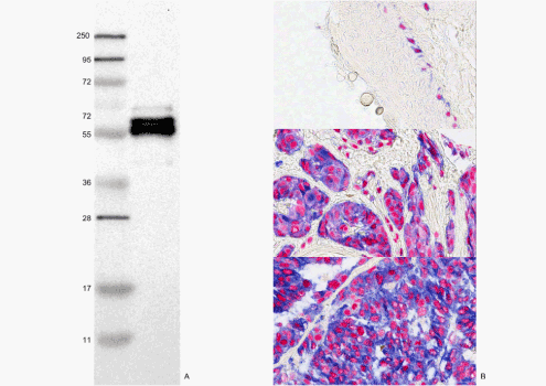
| Figure 2: Characterization of the MITF antibody (HPA003259). (A) A Western blot in SK-MEL-30 melanoma cell line demonstrating a doublet 55 and 60 kDa of the expected size where one band is phosphorylated (right lane). The left lane shows the molecular weight markers (250, 130, 95, 72, 55, 36, 28, 17 and 11 kDa) (B) A double immunostaining with Melan-A staining blue in the cytoplasm and MITF staining red in the nucleus. At the top benign melanocytes in the basal layer of normal skin, in the middle a benign melanocytic naevus and at the bottom malignant melanoma. (scale bar = 50μm). |