|
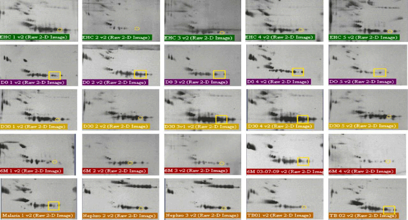
| Figure 2: 2DE map of total urine proteome: Proteins were separated by IEF in the neutral range (pH 3–10) followed by gradient 10% SDS–PAGE, and the resulting 2DE protein arrays were detected by staining with silver stain. Solid circles indicate absence of SSP 9101 while presence of this protein spot is indicated by square. EHC = endemic healthy controls; D0 = active VL patients (day-0); Dis = Patients after 30 days treatment; 6M = cured VL after 6 month follow-up; TB = tuberculosis patients. |