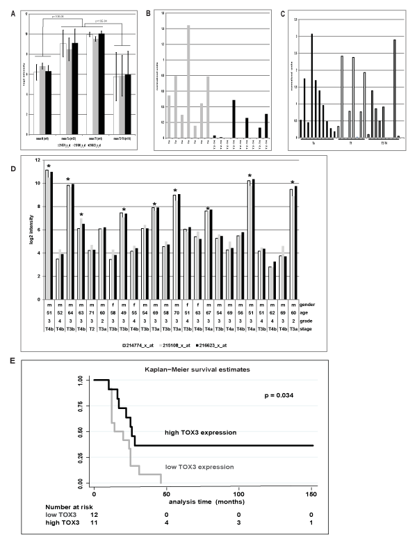
A) Log2 median expression values with standard deviation of normal urothelium (n=9), non-muscle invasive (n=56) and muscle-invasive tumors (n=24) analyzed on U133A microarrays with three different TOX3 probes.
B) RT-qPCR analysis of 16tumors previously analyzedon U133A microarrays confirmed a significant (p=0.02) upregulation of TOX3 in non-muscle invasive Ta tumors (n=7, median 0.542) compared to muscle invasive T2-4 tumors (n=9, median 0.032). Data were normalized to UBC.
C) TOX3 transcript localization in clinical samples assessed by ISH using a tissue microarrayshowed that the TOX3 transcript localized in the tumor cells.
D) TOX3 transcript expression shown as log2 intensities obtained from a panel of 24 T2-T4 tumor samples analyzed on U133A arrays with three probes for TOX3. Samples with high TOX3 expression log2>6 are labeled with an asterisks.
E) Kaplan Meier survival estimates on 23 of the 24 T2-T4 samples analyzed on U133A arrays with detailed patient follow up data. Preliminary data showed that patients with T2-4 tumors with high TOX3 levels showed a significant (p=0.034) better survival compared to those with low or absent TOX3 expression.