|
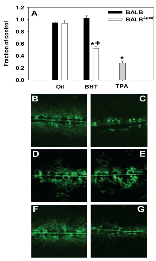
| Figure 3: Inhibition of GJIC after a 4 h exposure to BALF is both treatment and strain dependent. A) Graph of quantitated images from both BALB and BALBLps-d BALF-treated C10 cells following the SL/DT assay using Lucifer Yellow fluorescent dye. N=4-7 per treatment group, repeated twice. Data were normalized to the medium control and presented as mean + SEM for fraction of control. *, P<0.05 for BHT compared to oil treatment groups; +, p<0.05 for BALB versus BALBLps-d mice. More inhibition is evident in cells treated with BALF from BHT-treated BALBLps-d (G) compared to all other BALF treatments. (B) C10 cells treated with serum-deprived media alone as control; (C) C10 cells treated with TPA as a known inhibitor of GJIC. (D) C10 cells treated with BALF from oil-treated BALB mice; (E) C10 cells treated with BALF from BHT-treated BALB mice; (F) C10 cells treated with BALF from oil-treated BALBLps-d mice; (G) C10 cells treated with BALF from BHT-treated BALBLps-d mice. Magnification is 100X. |