|
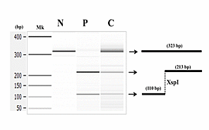
| Figure 2: Restriction enzyme digestion. Microcapillary electrophoresis of digested products. The PCR-amplified product spanning the exon 3 region was subjected to digestion with Xsp1. Left, products were separated via electrophoresis. Mk, DNA size markers. Right, product structures are displayed schematically. XspI, Xsp1 recognition site. In the normal genotype (N), the amplified product remained intact. In the patient (P), two small bands were visualized: a larger 213-bp product and a smaller 110- bp product. In our survey of 100 genomic DNA samples, one sample displayed three bands, indicating carrier (C) status |