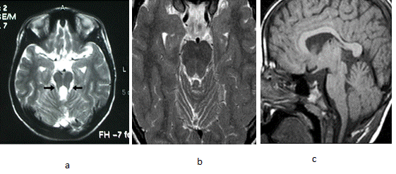
Figure 2:
Brain MRI, axial T2-weighted images at the level of the midbrain of Case 1 (a) and Case 2 (b). Sagittal T2-weighted image of case 2 (c).