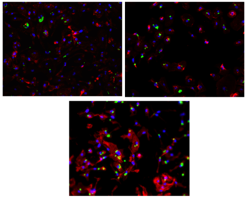
(a) The blue dots represent the DAPI stain of the nuclei, the red area is for the F-actin stain and the green dots represent the fluorescence stain of the yeast phagosomes (courtesy of the Dr. Annamaria Gal).
(b) The blue dots represent the DAPI stain of the nuclei, the red area is for the F-actin sain and the green dots represent the fluorescence stain of the yeast phagosomes (courtesy of the Dr. Annamaria Gal).
(c) The blue dots represent the DAPI stain of the nuclei, the red area is for the F-actin sain and the green dots represent the fluorescence stain of the yeast phagosomes (courtesy of the Dr. Annamaria Gal).