|
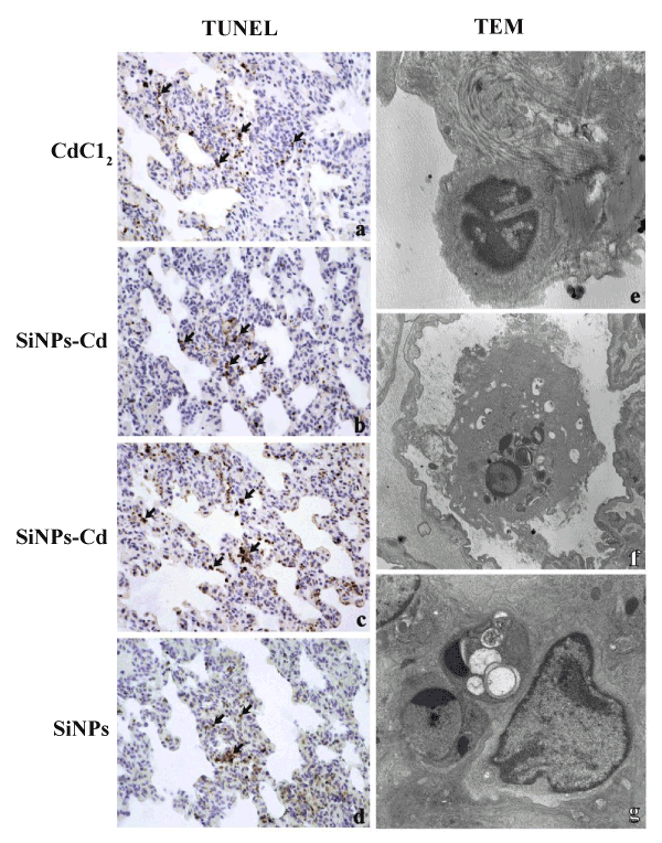
| Figure 4: (a-d) Representative micrographs showing apoptosis, detected by TUNEL staining, 24 hr (a-b) and 30 days (c-d) after i.t. exposure to CdCl2 (a), SiNPs-Cd (b-c) and SiNPs (d). TUNEL positive cells (chromatin condensation) detected in stromal and epithelial areas (a-d), in which labelledpneumocytes and macrophages (arrows) were observable. (eg) Electron micrographs showing different stages of apoptotic cell death: pyknosis (e), karyorhexis (f) and apoptotic bodies formation (g).Objective magnification: 40 x (a-d); Original magnification: x 12000 (f), x 7000 (e, g). |