 |
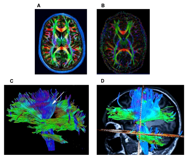
| Figure 3: DTI scans of the athlete 1 year after last concussion. Three-dimensional DTI images were acquired in the axial plane parallel with anterior and posterior commisure axis covering the entire brain (Siemens MAGNETOM Spectra 3T Skyra). A single shot spin-echo echo planar imaging (EPI) was used for DTI acquisition with the following parameters: 44 slices without a gap, FoV=230 mm, slice thickness=2 mm, TR=5900 msec, TE=95 ms, bandwidth=1000 Hz, echo spacing=1 ms.For whole brain coverage, the total DTI scan time was about 15 min. Fractional anisotropy (FA) map generated with syngo DTI Tractography for a healthy person (A) and a subject with concussion (B). White matter fiber tracts seen with syngo DTI Tractography (256 diffusion directions) in a healthy person (C) and a subject with concussion (D). |