 |
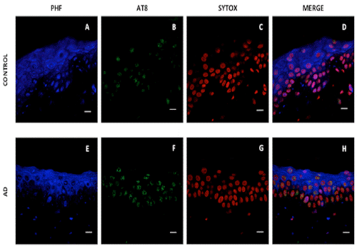
| Figure 4: Skin immunofluorescence. Confocal microscopy. PHF (Cy5) and AT8 (Alexa Fluor 488) antibodies. Control subject (A-D) and AD patient (E-H). Cell nuclei stained with Sytox (C-G). Positive juxtanuclear staining is observed in AD patient with AT8 antibody. Scale bar 10 μm. |