 |
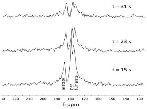
| Figure 7: Representative spectroscopy of hyperpolarized DES in a RENCA bearing animal. The first spectrum is taken 30 s after tail vein injection of 10 μmol of DES and uses a 30° flip angle. The tumor is placed in the center of solenoid coil and a small 13C-acetate phantom was placed next to the tumor as a reference. |