|
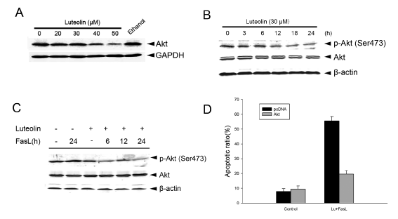
| Figure 3: The inactivation of Akt contributes to sensitized cell apoptosis. (A) HepG2 cells were treated with 20μM, 30μM, 40μM, 50μM luteolin for 16h. Cells lysates were detected by immunoblot analysis using indicated antibodies. (B) HepG2 cells were pretreated with luteolin as low as 30μM for 24h. Cells lysates were detected by immunoblot analysis using indicated antibodies. (C) HepG2 cells were pretreated with 30μM luteolin for 2h, followed by treatment with FasL (60ng/ml) for 3, 6, 12, 24 h. Cells lysates were detected by immunoblot analysis using indicated antibodies. The content of β-actin was used as a loading control. (D) HepG2 cells were transiently transfected with pcDNA and wt-Akt, after 24h, the cells were treated with luteolin (30μM × 24h) followed by FasL (60ng/ml × 22h). Cell lysate was used for Tunel assay. |