 |
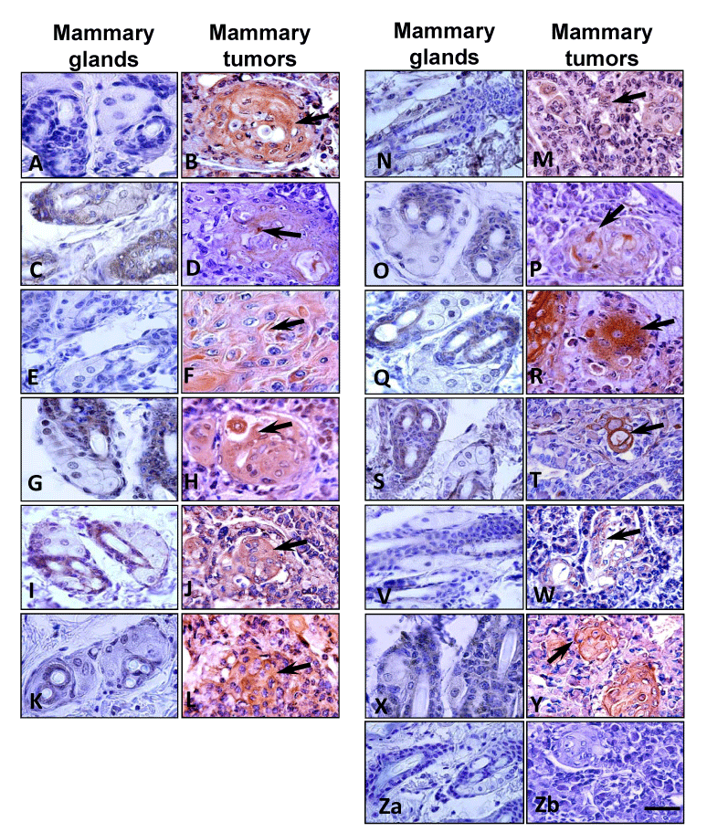
| Figure 2: Representative pictures from the immunohistochemical determination of several molecules expressed by mammary glands from mice treated with PEGLPrA2 and mammary tumors (DMBA-MT) developed in DIO-C (untreated mice), respectively. (A, B) AhR; (C, D) OB-R; (E, F) Leptin; (G, H) VEGFR-2; (I, J) IL-1R tI; (K, L) Notch1; (M, N) Notch2; (O, P) Notch3; (Q, R) Notch4; (S, T) JAG1; (V, W) Survivin; (X, Y) Bcl-2; (Za, Zb) Negative control. Similar results were obtained from mammary glands without MT from the same animal (not included). Arrows indicate positive staining in MTs. Bar = 200 μm. |