|
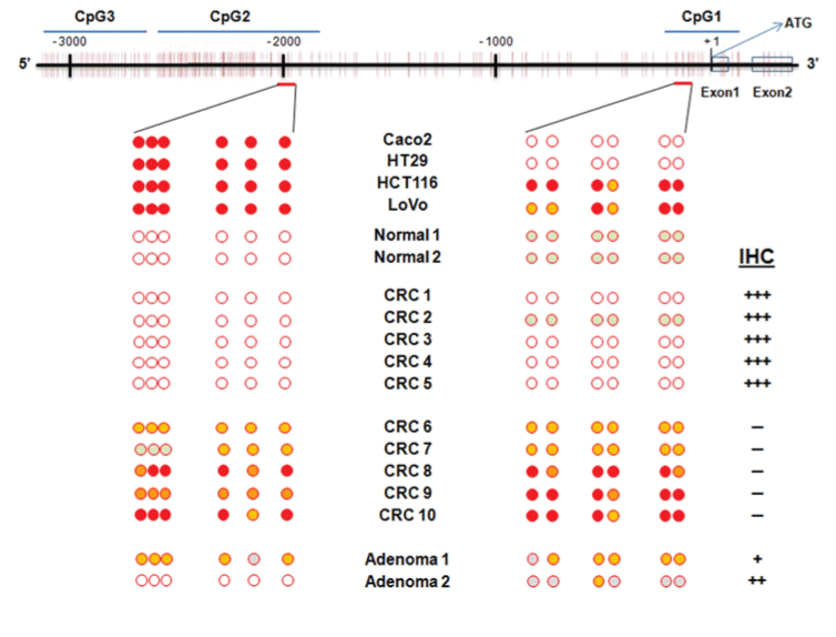
| Figure 6: Representative methylation analysis of CpG1 and CpG2 islands in the upstream region of μ-protocadherin gene in CRC cell lines (CaCo2, HT29, HCT116, Lovo), normal colorectal mucosa (Normal), carcinomas (CRC) and adenomas (Adenoma). CpG sites are indicated by vertical bars. The level of methylation observed at the analysed DNA region was graded as follows: 0-15 %, absence of methylation (white circle); 16-35%, low methylation (grey circle); 36-75%, moderate methylation (orange circle); 76-100%, high methylation (red circle). The level of μ-protocadherin protein expression arising from immune-histochemical analysis was indicated in the right of the figure. |