|
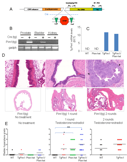
| Figure 1: (A) Scheme of the transgene strategy used. (B) Relative expression of PIM1 in Pim1/PSA-Cre mice after 2 rounds of hormone treatment. RNA was extracted from different tissues of 24 weeks old mice after testosterone and estradiol treatment. Reverse transcriptase PCR was performed to obtain cDNA, which was amplified using specifically designed primers. PCR fragment length was checked on a 1.5% agarose gel. (C) Levels of transgenic PIM1 mRNA determined by quantitative RT-PCR. Graph shows average levels of expression in the bladder of PIM1 mRNA of per genotype performed in triplicate. Data were normalized to the endogenous levels of GADPH in each sample. ND: Not detected. Transgenic PIM1 mRNA was not detected in WT or PTEN-Het mice. (D) Urothelial hyperplasia in hormone treated mice. To determine the development of urothelial hyperplasia due to hormone treatment, 8-week-old untreated mice of each genotype and hormone treated mice of corresponding genotypes, were sacrificed and bladder tissue was taken and distended with fixative (10% formalin). Upper pictures: Representative hyperplasia observed in bladder walls. Bottom pictures: Pictures show representative increases in bladder size over treatment course. H&E staining of bladders from tgPim1 mice before treatment and after 1 or 2 treatment rounds, respectively. All pictures were taken at the same magnification, (Panoramic viewer – ZeissE) Urothelial hyperplasia grades reached. (E) Grading of hyperplasia in the different conditions. Hyperplasia of epithelial cells in bladder, before and after hormone treatment, was graded using the following grading scale: bh-grade 0: normal (2-3 cell layers); bh-grade 1: slight hyperplasia (4 cell layers); bh-grade 2: slight/moderate hyperplasia (5-8 cell layers); bh-grade 4: moderate hyperplasia (9-10 cell layers); bh-grade 4: moderate/severe hyperplasia (11-12 cell layers); bh-grade 5: severe hyperplasia (>12 cell layers). See text for details. |