|
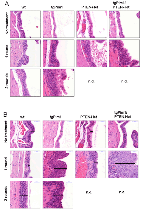
| Figure 2: Urothelial hyperplasia developed after hormone treatment. Example for (A) average of hyperplasia grade observed in each genotype (B) maximum of hyperplasia grade in each genotype. To determine the development of urothelial hyperplasia due to hormone treatment, 8-week-old untreated mice of each genotype and hormone treated mice (1 or 2 rounds) of corresponding genotypes were sacrificed and the bladder was taken. H&E staining of bladder tissue was used for grading and statistics as shown in Figure 1D. |