|
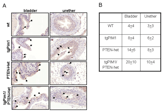
| Figure 4: p21waf1 nuclear stabilization in urothelial lesions. (A) Representative pictures. To examine p21waf1 in urothelial lesions, immunohistochemistry for p21 was performed in the bladder and ureter tissues of 16-week-old hormone-treated mice showing high grade and low-grade lesions. Picture shows p21waf1 staining in bladder and ureter lesions. Arrows show nuclear staining for p21waf1. (B) Average (± SD) percentage of nuclear p21 positive cells per genotype. |