|
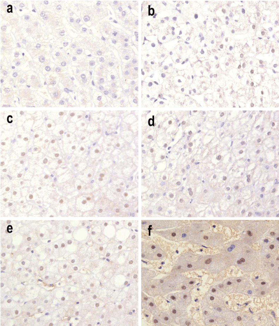
| Figure 1: Tissue sections showing the expression of c-MYC protein (nuclear staining) in liver parenchyma with CH in different fibrosis levels, which appears in brown colour. All pictures have magnification of 400x. a- there was no positive immunostaining of c-Myc (blue colour) in hepatocytes from NP; b- F0: cells with weak positive expression (+); c, d, and e- F1, F2, F3, respectively, as follows: cells with weak (+), with moderate (++), and f: F4, shows weak, moderate, and strong (+++) positive expression of c-MYC. |