|
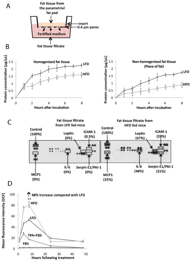
| Figure 2: Effect of high fat diet feeding on adipokine production and reactive oxygen species stimulation. A. Parametrial fat pad homogenate (0.5 g fat) from female SKH-1 mice that had been fed a 60% kcal high fat diet (HFD) or low fat chow diet (LFD) for 4 months was placed in 1.0 mL of MEM on a hanging well insert. Cytokines and other fat constituents passed through a polyethylene terephthalate membrane with 0.4 μm pores in the insert for 4 hours to provide fat tissue filtrate in MEM. B. Fat was isolated and fat tissue filtrate was made as described in (A). Graph demonstrates the amount of protein in MEM following culture with parametrial fat tissue homogenate or parametrial fat pad secretions from an intact piece of fat. C. Elevated amounts of monocyte chemoattractant protein-1 (MCP-1), interleukin-6 (IL-6), leptin, Serpin E1/plasminogen activator inhibitor-1 (PAI-1) and intracellular adhesion molecule-1 (ICAM-1) were released from the parametrial fat of mice fed a HFD compared with a LFD. D. Mean fluorescence intensity of the 2’, 7’-dichlorofluorescein (DCF) (excitation 488 nm; fluorescence 530 nm) +/- 15 of 5,000 events was measured. Fat tissue filtrate (50μg protein/mL), or TPA (10 ng/mL) induced ROS in JB6 cells. HFD fat tissue filtrate treatment stimulated the formation of ROS 48% more than LFD fat tissue filtrate as measured by DCF fluorescence at 8 hours **P<0.01. All data are mean ±SEM, Student t test (unpaired, two-tailed). |