|
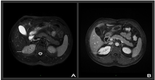
| Figure 1: Magnetic Resonance Imaging showed a small mass in the outside of the upper pole in the right kidney. A, Fat Suppression T2-weighted imaging showed a high signal mass measured 1.5- ×1.4-cm, which was not homogeneous inside the mass. B, Enhancement showed the mass was not enhanced. |