 |
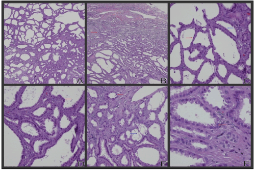
| Figure 2: The tumor is composed of small tubules and cysts, with some slender papilla pattern (A) and the boundary between tumor and normal tissue is ill-defined invasive. The red arrow showed the ill tumor’s boundary (B). Tumor cells are flat (red arrow) and cuboidal (blue arrow) with thin fibrous septa (C). Tumor cells also show high column (D, red arrow), hobnail cell (E, red arrow) or clear cytoplasm (E, blue arrow). Some tumor cells show prominent nucleoli (F, red arrow). |