|
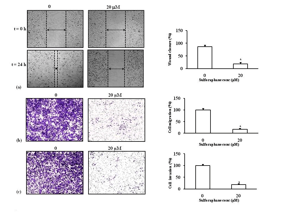
| Figure 5: Sulforaphane inhibits metastatic properties of breast cancer cells. (a) Wound healing assay was initiated by a uniform scratch in a pertidish containing MDA-MB-231 cells grown in confluence. Scratch was monitored under a microscope. The width of the scratch was measured and the percentage of closure was estimated. Results, expressed as mean ± SE (n=3) shows a significant inhibition of wound closure a(p<0.001) by sulforaphane. (b) For migration assay, MDA-MB-231 cells were seeded into the upper chamber of the transwell system. Cells were treated with suforaphane (20 μM) for 24 h and allowed to migrate. Migrated cells were fixed, stained with crystal violet and photographed. Acetic acid was used to extract cells bound to crystal violet. Optical absorbance was measured to quantitate the extent of cell migration, which was represented as percentage of migrated cells. Results are expressed as mean ± SE of three independent experiments. It was indicated that sulforaphane had an inhibitory effect on tumor cell migration. Extent of migration is distinctly different from the untreated cells, with significance level a(p<0.001). (c) Effect of sulforaphane (0, 20 μM) on the invasiveness of the metastatic breast cancer cells was studied using invasion assay. Treated cells were seeded in a transwell unit containing a membrane coated with ECM gel. Cells that have invaded through the ECM gel were fixed, stained with crystal violet and photographed. Quantification of invasiveness, as revealed by the absorbance of eluted crystal violet indicated reduction of invasive property of breast cancer cells. Bar diagrams represent percent of invaded cells. Results are expressed as means ± SE (n=3) and are significant compared to the untreated cells a(p<0.001). |