|
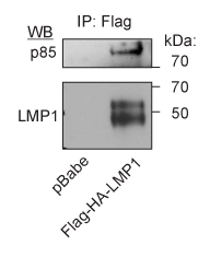
| Figure 2: Immunoisolation of LMP1-PI3K complexes. Rat-1 cells expressing FLAG-HA-LMP1 or vector only (pBabe) were grown to confluency and scraped into PBS. Cell pellets were frozen in liquid nitrogen, ground with a Qiagen TissueLyserII, and lysed in lysis buffer (20mM HEPES, 110mM Potasium Acetate, 2mM MgCl2, 250mM NaCl, 0.1% Tween, 1% Triton X-100). The lysate was spun at 18,000 x g for 5 min to pellet insoluble material and then incubated with Flag (sigma) antibody covalently bound to magnetic beads (Dyanal beads, Invitrogen) for 1h at 4 degrees. Bead-protein complexes were washed 5 times with lysis buffer then the proteins were eluted with a 0.5N NH4OH, 0.5mM EDTA solution and freeze dried in a speed vac. The protein pellet was dissolved in SDS sample buffer and separated by SDS-PAGE, transferred to nitrocellulose, and immunobloted for the p85 subunit of PI3K (upstate) and LMP1 (CS1-4). |