|
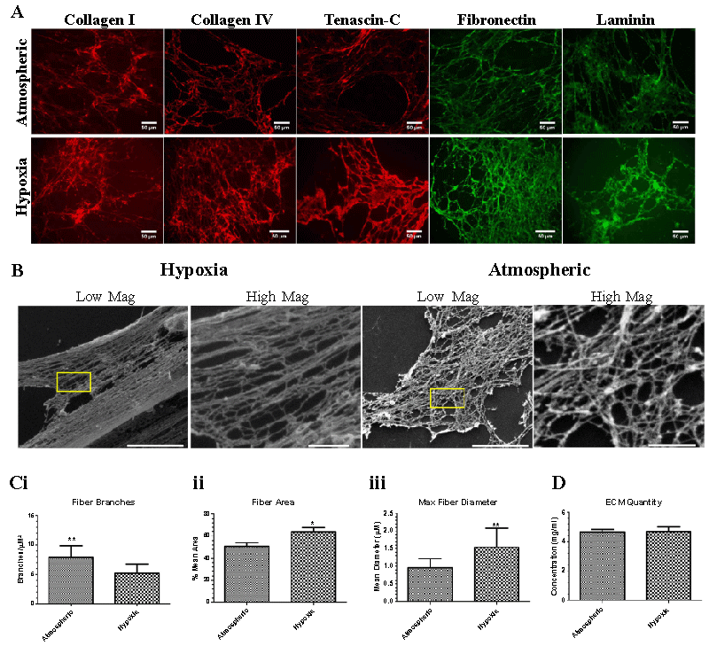
| Figure 4: Hypoxia altered the morphology and characteristics of de-cellularized ECM (A) ECM deposited from day 9 hypoxic and atmospheric-exposed cocultures was observed using immunofluorescence for fibronectin, collagens I and IV, laminin and tenascin-C. (B) SEM images depict the ultra-structural morphology of hypoxic and atmospheric ECM in low (left) and high (right) magnifications. Scale bars are 5μM and 1μM for low and high magnification images, respectively. (C) Quantification of fibers for differences in (i) branches, (ii) area, and (iii) maximum diameters in hypoxic and atmospheric ECM. (D) Total ECM concentration in hypoxic and atmospheric ECM. *p≤0.05, **p≤0.01, ***p≤0.001. |