|
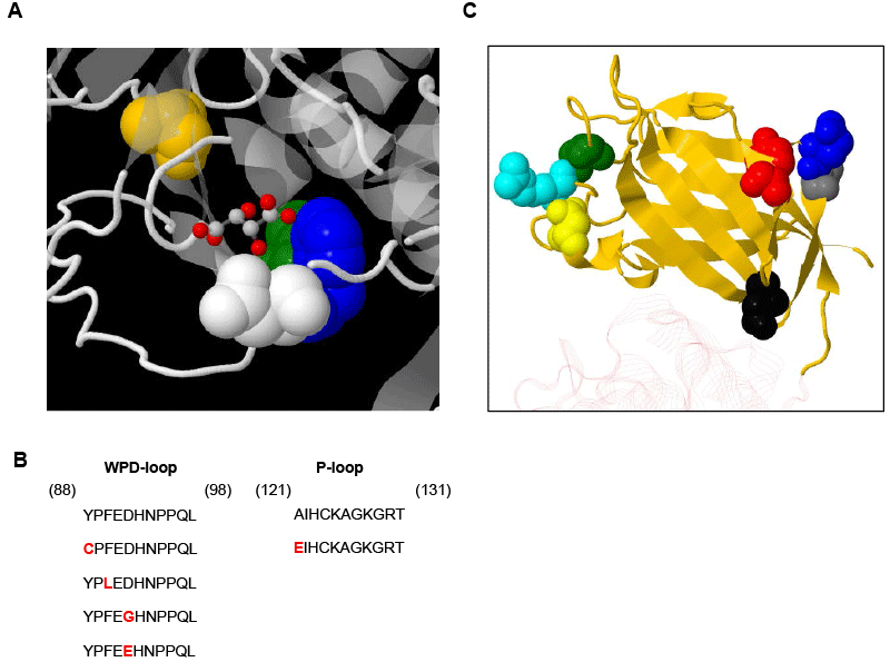
| Figure 3: Mutations of tumor suppressor PTEN in HNSCC. Crystal structure of PTEN protein is represented as ribbon diagrams. A. The functional active pocket of the phosphatase domain is shown. The mutated residues in the WPD-loop are highlighted in green (Y88), blue (F90), and white (D92). The mutated residue A121 is in yellow.B. Mutated residues Q245 (in blue) and R335 (in cyan) in the C2 domain are nonsense mutations. Y225, P246, and D252 are shown in red, gray, and black respectively. M205 (in green) is in the Cs1/2 loop; and D331G (in yellow) is in the C2α loop. C. Sequence of the WPD-loop and P-loop depicting the mutated residues within the walls of the active pocket present in the PTEN phosphatase domain. Mutated amino acid residues are in red. |