|
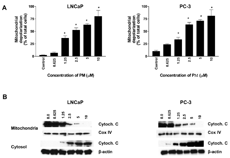
| Figure 3: PM induces mitochondrial depolarization and release of cytochrome c from mitochondria. (A) LNCaP and PC-3 cells were treated with PM at 0 to 10 μM for 20 h. Cells were loaded with mitochondrial potential sensor JC-1 (10 μg/ml) for 10 minutes at 22oC and analyzed by flow cytometry for cells fluorescing red (FL2 channel) or green (FL1 channel). Data are presented as percentage of cells with loss of mitochondrial potential difference. (B) Effect on mitochondrial cytochrome c. After treatment with PM (0 to 10 μM) for 20 h mitochondrial and cytosolic fractions were prepared from control and treated cells using ApoAlert Cell Fractionation Kit (Clonetech, Laboratories Inc., CA), and cytochrome c analyzed by western blotting. Data presented are from triplicate experiments. *p<0.05 compared to no PM controls. |