 |
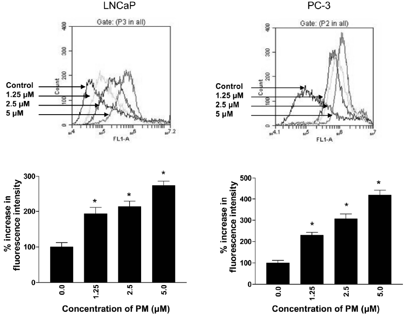
| Figure 4: PM induces intracellular ROS in prostate cancer cells. Subconfluent cultures of LNCaP and PC-3 cells were treated with PM (1.25 to 5 μM) for 2.5 h. Cells were reacted with 5 μM H2DCF-DA for 30 min at 37oC. Cells were collected and DCF fluorescence was measured by flow cytometry. Flow cytographs show shift in the mean DCF fluorescence intensity after treatment of LNCaP and PC-3 cells with PM. Bar graphs show percent increase in the mean DCF fluorescence intensity of cells treated with PM (Mean ± S.D. of three experiments) compared to untreated control cells (100%). *p<0.05 versus control. |