 |
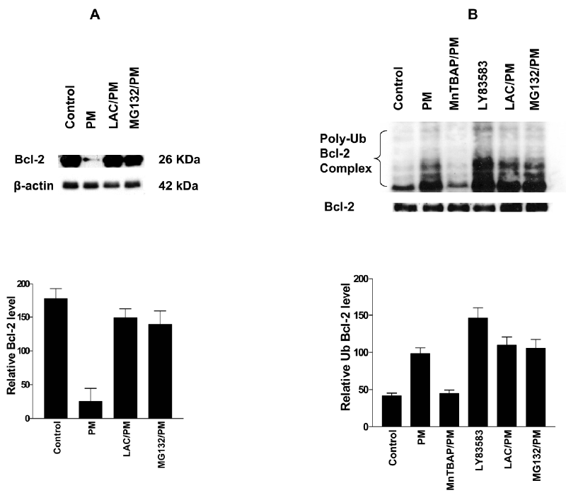
| Figure 7: Involvement of ROS in ubiquitination and proteasomal degradation of Bcl-2. (A) PC-3 cells overexpressing Bcl-2 were treated PM (5 μM) in the presence or absence of proteasome inhibitors LAC (10 μM) or MG132 (20 μM) for 20 h. Cell lysates were analyzed for Bcl-2 expression by western blotting. (B) PC-3 cells were treated with PM (5 μM) in the presence or absence of MnTBAP (100 μM), LAC or MG132. Cells were also treated with LY83583 alone (10 μM) for 6 h. Cell lysates were immunoprecipitated with anti-Flag antibody and immune complexes were analyzed for ubiquitin by western blotting. Ubiquitin was analyzed after treatment with PM for 6 h because ubiquitination was found to be maximal at this time point. Bar graphs show relative levels of Bcl-2 (A) and poly-ubiquitinated Bcl-2 (B). Similar results were obtained in two independent experiments. |