 |
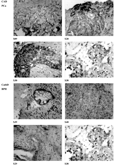
| Figure 4: Cathepsin D immunohistochemistry demonstrating the specific locations of CathD expressing cells in the neoplastic tissue. The expression was restricted to specific tissue sites within the tumor invasion, the positive cell populations were observed sparse in between the glandular tissue also showing that CathD plays an important role malignancy. The immunopositivity of this protein was restricted to specific tissue sites especially close to the base of the epithelium. It is important to note that S29 gave the highest immunopositivity, the expression level is higher than that observed in S39 and S40 but not as high as that observed in the control S38 (magnification X400). |