|
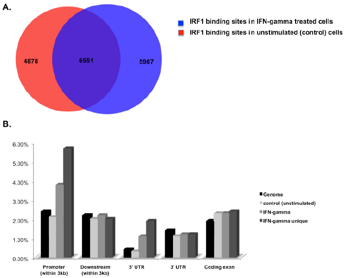
| Figure 1: ChIP-sequencing identifies IRF1 binding sites in H3396 breast cancer cells A. Shown are the number of peak regions identified in control (unstimulated) and IFN-gamma stimulated cells B. Positional chromosomal annotation (CEAS) of IRF1 bound peak regions represented as percent coverage. Shown is the set coverage for control, IFN-gamma unique (only found in IFN stimulated cells) and IFN-gamma total binding sites relative to the genome. |