 |
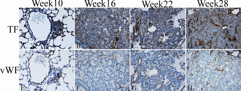
| Figure 2: IHC Detection of TF and vWF. Representative sections of IHC staining for TF and vWF, 40x magnification. All lesions contained cells that stained positively for TF and vWF. Week 10 sections (A, E) show a small tumor growing around a blood vessel. Weeks 16, 22, and 28 sections (B, C, D, F, G, H) are predominantly occupied by tumor. Positive TF staining is seen in the alveolar capillaries and macrophages. Intra-lesion heterogeneity of TF expression is also present. TF expression is greatest in the neovascular-rich tumor stroma. |