|
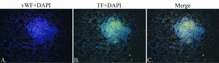
| Figure 4: Fluorescent Detection of vWF and TF in Urethane-Induced Lung Tumors. Representative section triple-stained immunofluorescently (IF) for vWF (Alexa 568), TF (tyramide-Alexa 488), and DNA (DAPI), 10x magnification. Staining for TF was stronger within the lesions and the strongest staining for TF was co-localized with vWF. (A) Vascular endothelial cells (VECS) and pericytes have a positive stain for vWF. Pre-existing pulmonary vessels are observed outside of the lesion, and developing microvessels are noted within the lesion. The vWF staining pattern indicates that the existing vessels may be forming extensions as tumor microvessels. (B) TF was observed in tumor-associated blood vessels. TF was expressed by normal lung tissue, including alveolar capillaries, while bronchiolar epithelial cells (BECS) showed only weak staining for TF. (C) Expression of TF by tumor cells was highest in regions of active angiogenesis. |