|
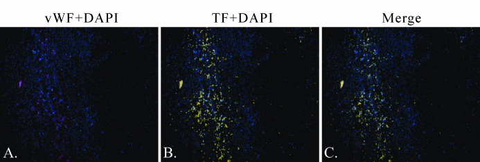
| Figure 7: Fluorescent Detection of vWF and TF in Urethane-Induced Lymphoma. Representative sections triple-stained immunofluorescently (IF) for vWF (Alexa 568), TF (tyramide-Alexa 488), and DNA (DAPI), 10x magnification. (A) Blood vessels and areas of angiogenesis are present. (B) TF staining is strong within the tumor stroma but absent inside the tumor. (C) The strongest staining for TF is co-localized with vWF. vWF appears to be a subset of TF. |