 |
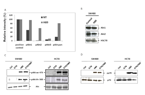
| Figure 2: H89 induces phosphorylation of Akt on serine 473 and threonine 308 and the mTOR target kinase, p70S6K. (A) SW480 cells were treated for 48 hours with H89 (10 μM) or left untreated (NT). Cell lysates were analyzed with a phospho-MAPK Array kit to detect the phosphorylation status of Akt isoforms (Akt1, Akt2, Akt3). Each point was analyzed with image J software to express the results in guise of relative intensity. The positive control is an internal control supplied with the kit and represents 100% of the signal (pixel density). (B) Western blot analysis of Akt1 and Akt2 in SW480 cells after H89 treatment (10 μM) for 48 hours. HSC70 was used as a control for equal protein loading expression (1 representative of 3 independent experiments). (C and D) SW480 cells (3 × 10⁵/mL) and HCT8 cells (3 × 10⁵/mL) were treated with GTN (10 μM) or/and H89 (10 μM) or left untreated (Ctrl) for 24 hours at 37°C. Total proteins were obtained and subjected to western blot analysis for Akt, pAkt ser 473, pAkt thr 308 expression (C), p70 and pp70 expression (D) (1 representative of 3 independent experiments). Results have been obtained after a short exposure of the film (10 min). However, longer exposure of the film (30 min and more) permits the visualization of a slight expression of pAkt1 in non treated cells (data not shown). |