|
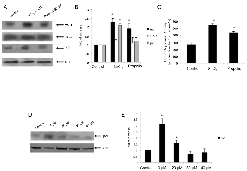
| Figure 3: (A) Western Blot analysis of Hmox1, Hmox-2 and p21 following treatments for 72 h with Chilean propolis ethanolic extract (80 μg/ml) and 10 μM SnCl2. (B) Densitometric analysis of western Blot following actin normalization. (C) Hmox activity following different pharmacological treatment. (D) p21 protein expression following treatments with tricarbonyldichlororuthenium (II) dimer (CORM-II) at different concentrations (5-50 μM). (E) Blots shown are representative of Western blot analysis from three separate experiments (*p<0.001 vs control). |