|
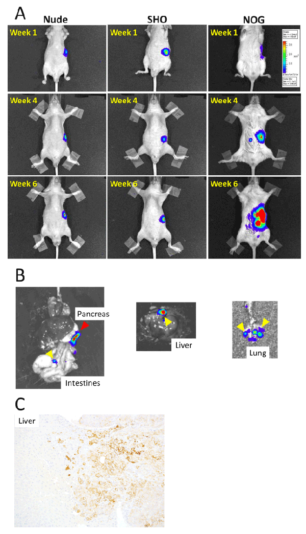
| Figure 2: (A) In vivo imaging of an orthotopic implantation model of pancreatic cancer in Nude (left), SHO (center), and NOG (right) mice. In Nude and SHO mice, the size of the luminescence from tumors implanted in the pancreas remained unchanged during the experimental period. On the other hand, the luminescent signal in NOG mice expanded over the week. (B) Ex vivo imaging of internal organs of the NOG mouse showed bioluminescence signals in the pancreas (left; red arrowhead), intestines (left; yellow arrowhead), liver (center; yellow arrowhead), and lungs (right; yellow arrowheads). (C) The immunostaining of HLA class I-A,B,C clearly highlighted the nest of human cancer cells in the liver of the NOG mouse. Original magnification, ×200. |