|
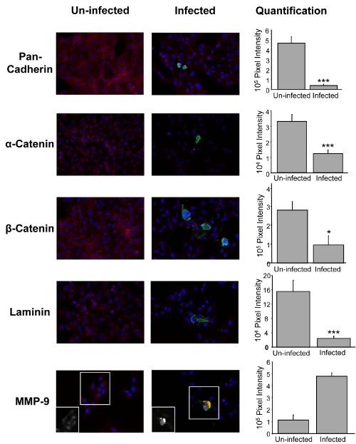
| Figure 6: Infection of A549 cells by F. novicida induces the degradation of adherens junction and basal membrane proteins. A549 cells were infected for 24 hrs with F. novicida (U112) and fixed for immunofluorescence staining in order to determine the expression of adherent junction proteins pan-cadherin, α-catenin and β-catenin, basal membrane protein laminin, matrix metalloproteinase 9 (MMP9) and U112. U112 was stained by using an antibody against its LPS (green). Nuclei of cells (blue) were visualized by staining with DAPI. The first column has representative images of un-infected cells. The second column depicts infected cells. MMP9 images have an inset in black and white that corresponds to MMP9 staining alone. The third column depicts the quantification of the expression of the different proteins analyzed. Quantification was performed by using IPlab software; the graphed data represents an average of 15 single cells per protein and is given as pixel intensity. One asterisk, P < 0.05; two asterisks, P < 0.05; three asterisks, P < 0.0005. |