 |
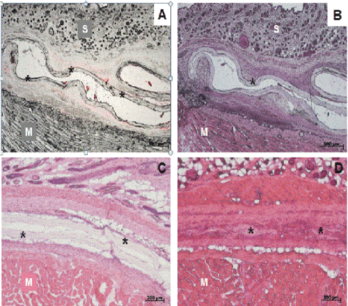
| Figure 4: Histological evaluation of extracted tissues. Consecutive cryostat section one week after surgery (top row) showed seeded CCC (*) lying folded between skin and rectus muscle. Overlay of unstained section (A) demonstrated red fluorescent PKH26-labelling of seeded HUC. HE-stained section (B-D) revealed a continuous and intact matrix one (B) and two weeks post-op (C). After four weeks ongoing degradation process of CCC could be documented. Note: * = CCC, M = muscle, S = skin. |